
A Look into the Eye's Biomechanics
Authors: Padmanabhan, P., Lopes, B.T., Eliasy, A., Abass, A., and Elsheikh, A.
Journal: Current Eye Research
Publication Date: Apr 2022
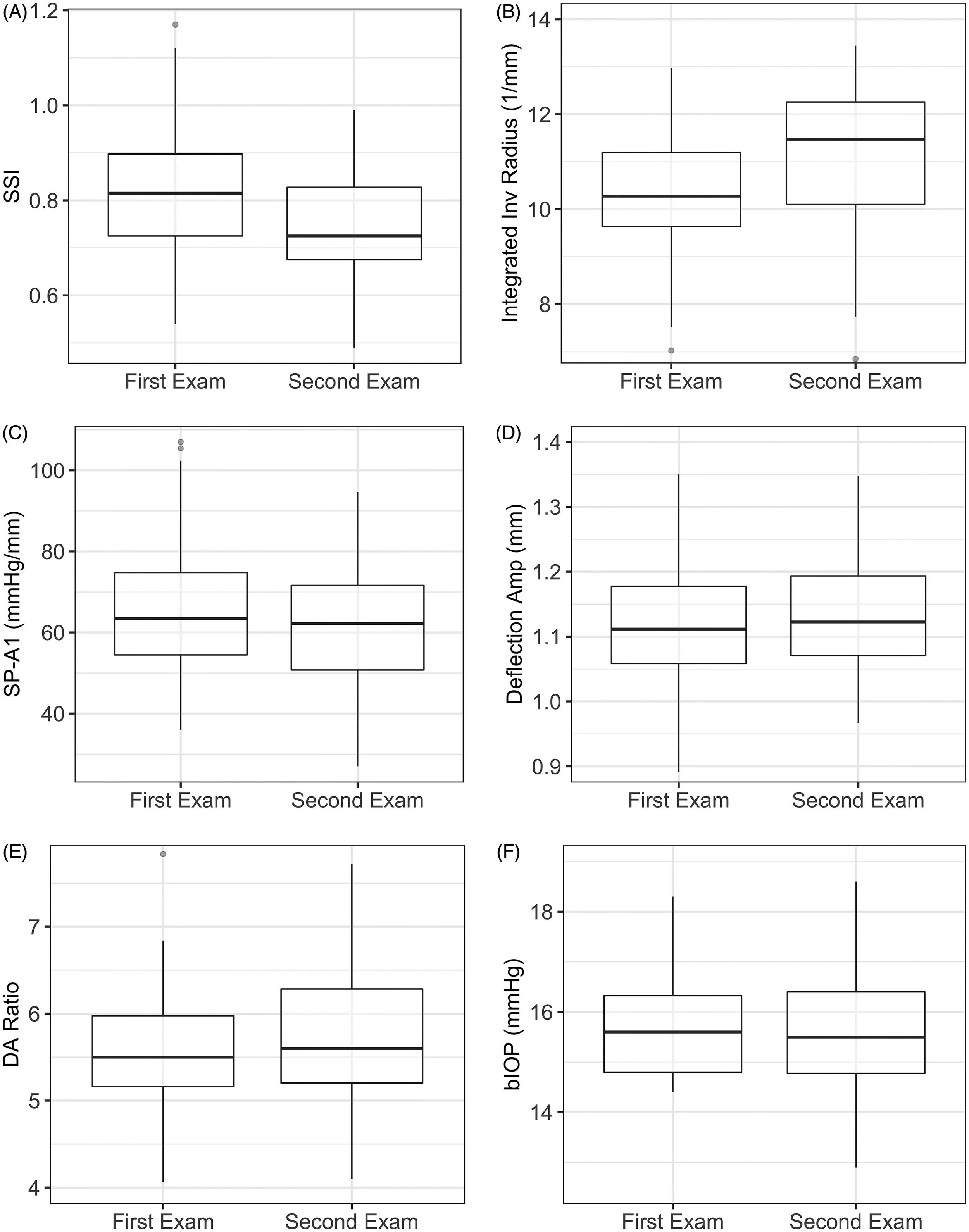
Distribution of the corneal biomechanical parameters at the first and last examinations. A: SSI: stress-strain index. B: Integrated Inv Radius: integrated inverse radius. C: SP-A1: stiffness parameter at the first applanation. D: Deflection Ampl: deflection amplitude. E: DA Ratio: deflection amplitude ratio. F: bIOP: biomechanically corrected intraocular pressure.
Summary:
Keratoconus is an eye condition that affects the cornea, leading to vision problems and a reduced quality of life. To better understand the progression of this disease, our research team examined the biomechanical changes associated with it using Corvis ST. By studying 32 cases of progressive keratoconus, we were able to identify significant deterioration in corneal stiffness and other biomechanical parameters.
Our findings revealed that the Stress-Strain Index (SSI) and Integrated Inverse Radius (IIR) showed the most significant changes between examinations, suggesting that they could be used as reliable markers of keratoconus progression. The SSI, a recently introduced in vivo estimation of corneal biomechanical status, was particularly notable for its small overlap between the two sets of results.
We also observed trends toward corneal softening in other parameters, such as the Stiffness Parameter A1 (SP-A1) and Deflection Amplitude (DA). While these measurements were not as sensitive as the SSI and IIR, they still provided valuable insights into the biomechanical changes taking place in the cornea during keratoconus progression.
In conclusion, our research demonstrates that in vivo measured biomechanical deterioration occurs with keratoconus progression. The SSI and IIR, in particular, show promise as potential biomarkers for the disease's progression. Further studies are needed to confirm these findings and to explore the use of these parameters alongside other recently developed indicators, such as the Corneal Biomechanical Factor (CBiF).